ECR 2018 / C-2418
Decoding the Contrast Enhanced Brain
Congress:
ECR 2018
Poster Number:
C-2418
Type:
Educational Exhibit
Keywords:
Neoplasia, Infection, Education and training, Education, Contrast agent-intravenous, MR, Neuroradiology brain, Contrast agents
Authors:
V. B. Pai, K. Joshi, K. Gupta, C. Trivedi, A. Agrawal, B. Pai, R. Kharche, D. Lokhande; Mumbai/IN
DOI:
10.1594/ecr2018/C-2418

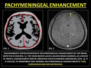
Fig. 6:
REPRESENTATIVE IMAGES OF PACHYMENINGEAL ENHANCEMENT

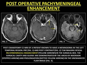
Fig. 7:
POST OPERATIVE PACHYMENINGEAL ENHANCEMENT

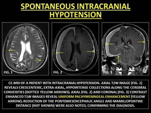
Fig. 8:
SPONTANEOUS INTRACRANIAL HYPOTENSION

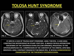
Fig. 9:
TOLOSA HUNT SYNDROME

Fig. 10:
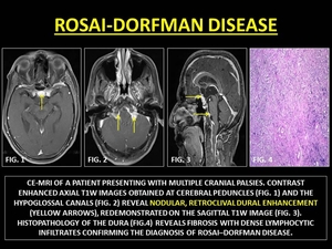
ROSAI-DORFMAN DISEASE

Fig. 11:
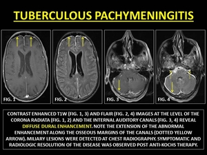
TUBERCULOUS PACHYMENINGITIS

Fig. 12:
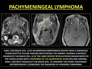
PACHYMENINGEAL LYMPHOMA

Fig. 13:
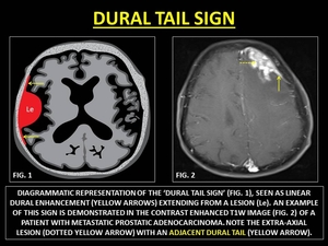
REPRESENTATIVE IMAGES OF 'DURAL TAIL SIGN'

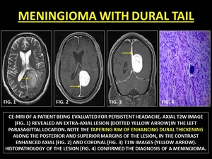
Fig. 14:
MENINGIOMA WITH DURAL TAIL

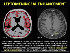
Fig. 15:
REPRESENTATIVE IMAGES OF LEPTOMENINGEAL ENHANCEMENT

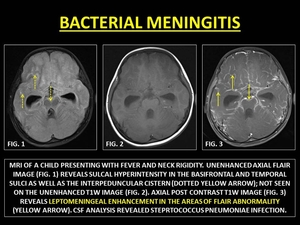
Fig. 16:
BACTERIAL MENINGITIS

Fig. 17:
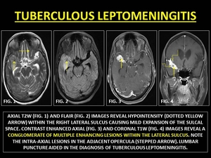
TUBERCULOUS LEPTOMENINGITIS

Fig. 18:
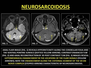
NEUROSARCOIDOSIS

Fig. 19:
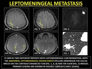
LEPTOMENINGEAL METASTASIS

Fig. 20:
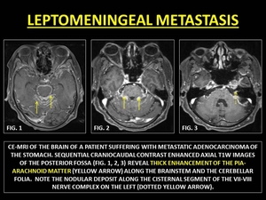
LEPTOMENINGEAL METASTASIS

Fig. 21:
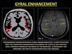
REPRESENTATIVE IMAGES OF GYRAL ENHANCEMENT

Fig. 22:
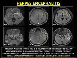
HERPES ENCEPHALITIS

Fig. 23:
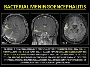
BACTERIAL MENINGOENCEPHALITIS

Fig. 24:
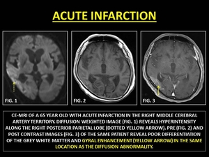
ACUTE INFARCTION

Fig. 25:
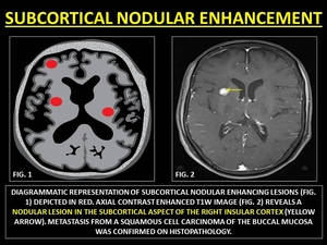
REPRESENTATIVE IMAGES OF SUBCORTICAL NODULAR ENHANCEMENT

Fig. 26:
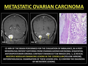
METASTATIC OVARIAN CARCINOMA

Fig. 27:
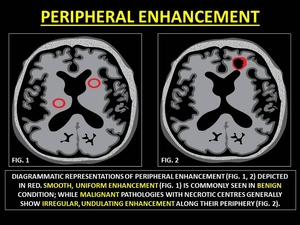
REPRESENTATIVE IMAGES OF PERIPHERAL ENHANCEMENT

Fig. 28:
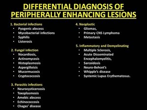
DIFFERENTIAL DIAGNOSIS OF PERIPHERALLY ENHANCING LESIONS

Fig. 29:
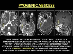
PYOGENIC ABSCESS

Fig. 30:
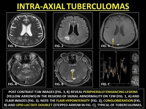
INTRA-AXIAL TUBERCULOMAS

Fig. 31:
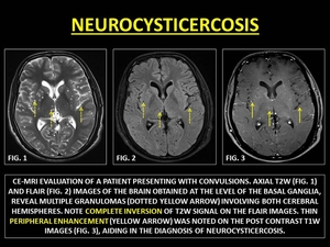
NEUROCYSTICERCOSIS

Fig. 32:
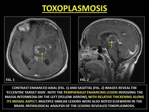
TOXOPLASMOSIS

Fig. 33:
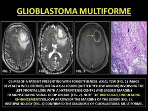
GLIOBLASTOMA MULTIFORME

Fig. 34:
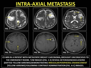
INTRA-AXIAL METASTASIS

Fig. 35:
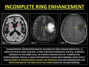
INCOMPLETE RING ENHANCEMENT

Fig. 36:
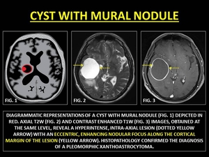
CYST WITH MURAL NODULE
 CONTRIBUTED BY DR. PRASHANT MUDGAL.")
Fig. 37:
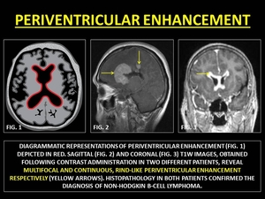
PERIVENTRICULAR ENHANCEMENT

Fig. 38:
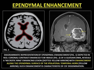
REPRESENTATIVE IMAGES OF EPENDYMAL ENHANCEMENT

Fig. 39:
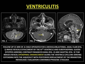
VENTRICULITIS

Fig. 40:
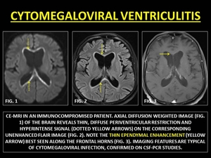
CYTOMEGALOVIRAL VENTRICULITIS

Fig. 41:
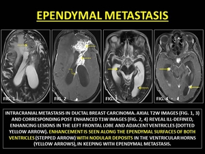
EPENDYMAL METASTASIS